
Author: Nikki Abela / Questions: Nikki Abela / Codes: / Published: 06/02/2018
Doctor, can you cast your eyes over this ECG for me?
It’s a phrase you’ll come to know well. Especially, you’ll find, if you’re sitting at a certain desk or in a certain area. You’re more likely to be given adult ECGs to view, but if you’re interested in learning more about paediatric ECGs, visit our blog here.
It’s a phrase which at first may fill you with dread, but with a simple system, you will find you will soon become expertly efficient at picking out obvious problems.
Never say no unless you are in the middle of something important and stopping for a few minutes will be detrimental – it is important that we support whoever is tasked with needing to ask for ECGs to be checked and this is a great learning opportunity for you as you will always have someone to ask if you’re not sure. Do remember though, to look at the ECG properly, and not to trust the computer read out!
This “safety-net” of getting doctors to check ECGs is a bone of contention for many senior doctors as it takes up a fair bit of time – but that is not for you to worry about, but it’s so important that in some trusts, only senior doctors can do this.
The system of checking ECGs is primarily there to make sure we don’t miss that all important STEMI that needs urgent transfer for primary PCI. But if you’re only looking at the ST segments on ECGs in this “quick look”, you’re definitely missing a trick.
Always ask or check the triage sheet for the presenting complaint – it will give you an idea of what you should be looking out for. In a patient with chest pain, for example, those ST segments are all the more important, in a patient who has taken an overdose, think about checking the QT and QTc, and in a patient with suspected sepsis, that new fast AF may be even more important.
Look at the Rate
It’s so basic isn’t it? I’m not going to go into how to read ECGs medical school style though – if it’s fast and they are in an irregular rhythm, or even regular (SVTs, for example), ask for a monitored bed and cast your eye over the patient. It’s likely they need prioritising, either to have their rate/rhythm dealt with, or the underlying cause treated (think of sepsis, for example, where there is some good evidence that patients may not benefit from rate or rhythm control). In this time of departmental crowding, monitored beds may be hard to come by – cast your eye over the patient, and make sure the coordinating senior knows about the patient, so they can juggle beds as needed.
It may be slow, in which case they are even more likely to need a cardiac monitor, unless they are the “goes to the gym regularly” prototype. These patients are likely to need to be in resus, on the defibrillator, especially if they have adverse features (shock, syncope, chest pain or symptoms of heart failure). Ask for senior input early.
The rate may be normal, and so may the rhythm, but they may still have worrying features – like heart block greater than the first degree or trifascicular block (1. Prolonged PR, 2. Left anterior or posterior fascicular block, 3. RBBB, in case you were wondering) – so they are going to need that monitored bed and alarms should be going off in your head if their triage note says “collapse ?cause”.
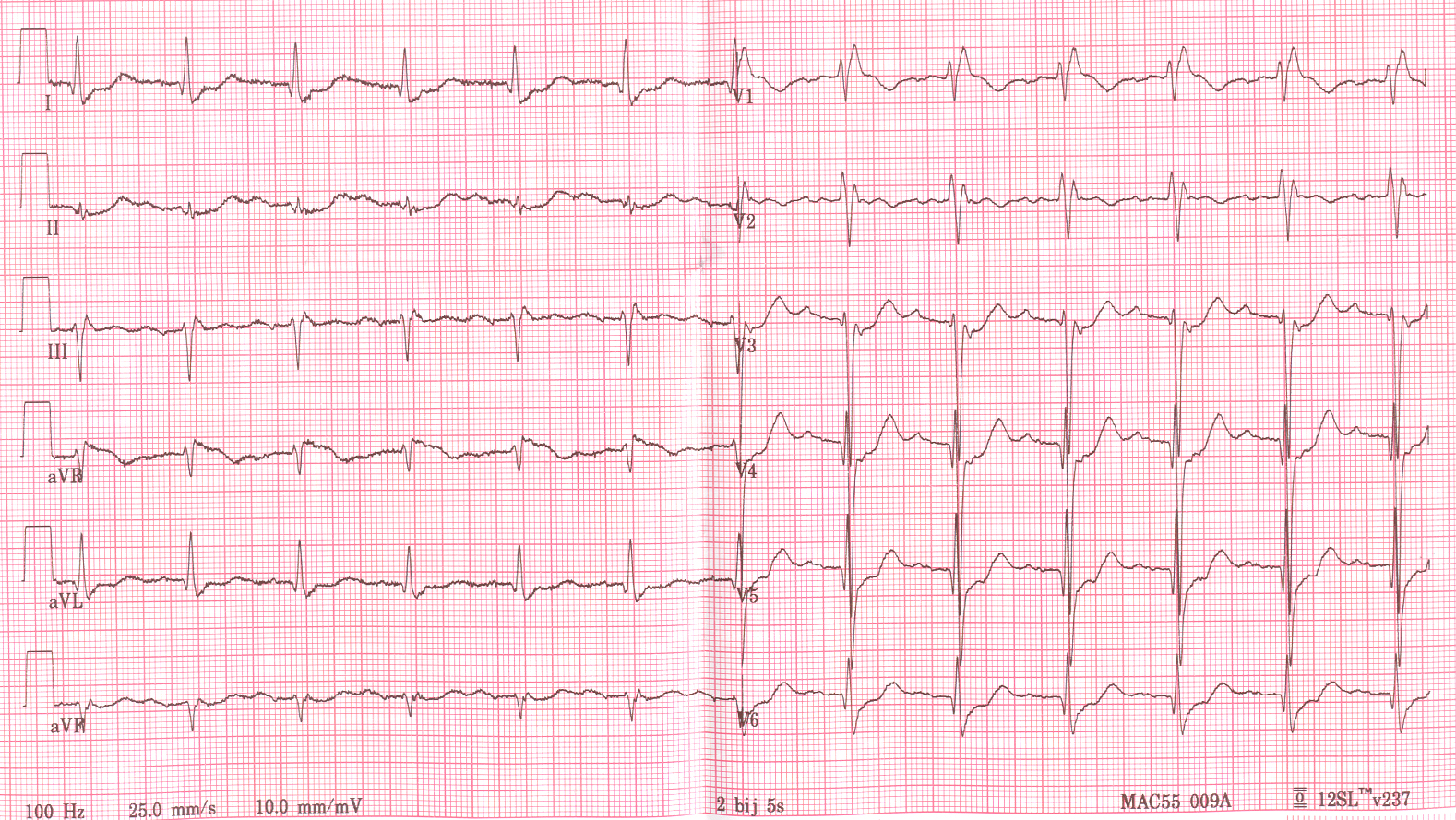
Trifascicular block.
Check the QT and QTc
Sadly, we see a fair share of overdoses in the ED and when an ECG is shoved under our noses, we may not be aware of the presenting complaint. Most drugs and electrolyte deficiencies prolong the QT and QTc, digoxin and hypercalcaemia shorten it. If it’s prolonged, especially in an overdose or patient with electrolyte deficiencies, this needs to be picked up for them to be treated asap. If the patient’s rate is fast, don’t trust the computer calculation of the QTc.
And of Course, Check Those STs
Yep, the ST segments have started the trend for checking ECGs, so don’t neglect them. If they are elevated, go to see the patient straight away. Remember, time is myocardium so the quicker they get that PPCI, the better their outcome. Don’t know how to get them this? Your hospital will have a policy, I’m sure.
Remember, that not everything that doesn’t look like a STEMI isn’t, and you need to know your atypical STEMI patterns and STEMI equivalents (do check the hyperlink for a super blog by emDOCs). If you’re seeing a strange ST pattern, don’t be afraid to google what it looks like if it reminds you of something but you can’t remember what. I have often found myself googling “sinusoidal T-waves” to jog my memory about Wellen’s syndrome, or looked up Brugada syndrome, if I’m worried my patient’s ECG looks somewhat similar.
Life in the Fast Lane has an excellent ECG library that is very handy to reference. If you haven’t come across it by now, you’re going to kick yourself for spending medical school without it. If you want to scare yourself, look at their “Killer ECG patterns” post!
If the ECG just looks ischaemic, and the patient is in pain, do go and give them analgesia and get them prioritised – just because they aren’t going for PPCI, doesn’t mean they aren’t having an MI.
When in Doubt, Check the old ECG
If you have easy access to old ECGs, do compare worrying signs with old ones as generally speaking, if they were there before, you likely don’t need to worry as much. We should probably all give patients copies of their ECG, especially if they’re abnormal. They can take a picture on their smart phone, then any doctor can easily compare ECGs. When you see a patient who doesn’t have an old ECG to compare to, let that be a reminder to give them a copy!
If the ECG Analyser says normal sinus rhythm (NSR) – it probably is
There’s an old adage that we shouldn’t look at the analyser comments on ECG as it will bias our interpretation. However, emerging evidence shows that if it says NSR, it probably is NSR.
That is, until it isn’t. You don’t want to be the exception to this rule, so if you follow these simple steps, it is likely that you will be safe.
Remember, when someone asks you to “cast your eye over an ECG”, it is not to check for the small nuances on the tracing, but rather to pick up issues that need prioritising. So don’t sweat the small stuff, use this exercise as a valuable tool to become excellent at spotting the “sick ECG” – if in doubt, cast an eye on the patient, and speak to a senior.