
Author: Richard Freeman / Editors: Gareth Lewis / Reviewer: Jason Kendall, Richard Freeman / Codes: / Published: 17/03/2021
Neonates can present with normal physiology to the paediatric emergency department. Studies have suggested that 1.9% of all patients present within the first month of life (1).
The most common diagnosis are the normal newborn (33.9%), indirect hyperbilirubinaemia (13.2%) and colic (5.8%)1. As such, a large proportion of these patients could be discharged without the need for paediatric review.
During this session we will explore common benign conditions that could be managed by the emergency physician with relevance to the published evidence and national guidelines.
In this first part we will concentrate or three key areas:
1) Benign skin conditions
2) Physiological jaundice
3) Lumps and bumps
Skin rashes:
1) Erythema Toxicum Neonatorum
2) Transient Pustular Melanosis
3) Millia
4) Harlequin Colour Change
5) Seborrhoeic Dermatitis
6) Mongolian Blue Spot

1) Erythema Toxicum Neonatorum

This is a common skin condition that affects 40-70% of all neonates2.
20% of cases present at birth however, most develop 24 to 48 hours later3.
Histologically the condition is characterised by collections of eosinophils just beneath the stratum corneum. However, the aetiology remains unknown3.
2) Transient Pustular Melanosis
![]()
This condition is not as common as Erythema Toxicum occurring in 1% of all Caucasian newborns5.
The condition is five times more prevalent in the Afro-Caribbean population
3) Milia

A very common condition that occurs in 50% of newborns7.
Histologically the condition is characterised by retention of keratin within the dermis.
4) Harlequin colour change

A condition that occurs in 10% of newborns8. However, it is often un-recognised.
The condition is thought to be related to hypothalamic immaturity resulting in dilatation of the peripheral vasculature.
5) Seborrhoeic Dermatitis

Occurs in infants between the second week of life and 6 months.
The exact cause is unknown however, there is evidence of involvement of the yeast Pityrosporum Ovale10.
6) Mongolian Blue Spot

Bilirubin metabolism

Unconjugated bilirubin (indirect bilirubin) is created from red blood cell breakdown and transported in the bloodstream mostly bound to albumin.
Unconjugated bilirubin is transported into the hepatocytes and conjugated with glucuronic acid.
Conjugated bilirubin (direct bilirubin) is excreted into the biliary system and, ultimately, the small intestine.
Most bilirubin is excreted into the stool but a small proportion is reabsorbed as part of the entero-hepatic circulation via the portal vein.
Neonates are more prone to jaundice as:
1) They have a relative polycythaemia and thus, increased red cell breakdown.
2) The liver is relatively immature and thus, unable to cope with normal bilirubin metabolism.
3) Changes in intestinal flora alter the entero-hepatic circulation.
In most babies jaundice is self-limiting and harmless.
This is termed physiological jaundice.
Physiological jaundice is very common with 60% of term and 80% of preterm (<37 weeks) infants developing jaundice in the first few weeks of life13
Bilirubin metabolism
Unconjugated bilirubin (indirect bilirubin) is created from red blood cell breakdown and transported in the bloodstream mostly bound to albumin.
Unconjugated bilirubin is transported into the hepatocytes and conjugated with glucuronic acid.
Conjugated bilirubin (direct bilirubin) is excreted into the biliary system and, ultimately, the small intestine.
Most bilirubin is excreted into the stool but a small proportion is reabsorbed as part of the entero-hepatic circulation via the portal vein.
Neonates are more prone to jaundice as:
1) They have a relative polycythaemia and thus, increased red cell breakdown.
2) The liver is relatively immature and thus, unable to cope with normal bilirubin metabolism.
3) Changes in intestinal flora alter the entero-hepatic circulation.
1) Erythema Toxicum Neonatorum

Typically the rash presents as a maculo-papular or vesiculo-pustular eruption over the face, trunk and limbs. Each lesion is surrounded by an area of erythema.
The palms and soles remain unaffected.
The child remains systemically well.
If the child is sick other differentials for vesiculo-pustular rashes in the neonate should be considered (i.e. HSV, candida, Staphylococcus)4.
2) Transient Pustular Melanosis
![]()
Typically the rash presents as a vesiculo-pustular eruption that can occur anywhere on the body, including the palms and soles.
Unlike Erythema Toxicum the lesions have no surrounding erythema and rupture to leave a characteristic pigmented macule.
The child remains systemically well
3) Milia
Milia presents as 1-2mm pearly white or yellow papules that typically occur on the face (especially the forehead, cheeks, nose and chin).
However, they may appear on any part of the body include the mucous membranes, palate or penis.
The child remains systemically well
4) Harlequin colour change
Harlequin colour change occurs when the newborn lies on their side. The dependant side develops erythema whereas the contralateral side blanches.
The colour change can last between 30 seconds to 20 minutes and resolves with crying or increased activity
5) Seborrhoeic Dermatitis
The condition typically presents with erythema to the face, neck and flexural folds and greasy scales to the scalp (i.e. cradle cap). The napkin area can also be affected.
Unlike atopic eczema (which tends not to present in the neonatal period) pruritus is uncommon9.
The child is systemically well and, although often distressing for the parents, unaffected.
Generalized seborrhoeic dermatitis accompanied by failure to thrive and diarrhoea should prompt an evaluation for immunodeficiency11
6) Mongolian Blue Spot
They present as dark blue-grey lesions typically involving the lumbosacral region.
They are present from birth.
The child remains systemically well
What is physiological jaundice?
Jaundice developing after the first 24 hours.
Jaundice resolving by 14 days of life in term (37-42 weeks) or 21 days in preterm neonates (<37 weeks).
The child is systemically well and thriving.
The total serum bilirubin is less than the treatment threshold (see below).
Otherwise, pathological causes should be considered.
1) Jaundice developing within the first 24 hours
Jaundice developing within the first 24 hours of life is likely to be related to a haemolytic process (i.e. ABO incompatibility) and thus, always pathological.
In young babies indirect bilirubin can pass the blood brain barrier and high levels can lead to long-term damage to the CNS (kernicterus).
Hence, such patients should have urgent serum levels taken and referred urgently to paediatrics.
2) Jaundice not resolving by 14 days of life in term (37-42 weeks) or 21 days in preterm neonates (<37 weeks)
Symptoms continuing beyond this period are termed prolonged jaundice and can be a clue to serious underlying liver disease such as biliary atresia.
As such infants require investigation they should be referred to paediatrics.
NB: such patients are often referred from the community and investigated in a specific prolonged jaundice clinics. We would recommend following local guidelines.
3) If the child is unwell or failing to thrive
If the child is unwell alternative diagnoses such as sepsis, dehydration or inborn errors of metabolism should be considered. The management of these conditions is beyond the scope of this module.
As a general rule of thumb:
- It is normal for a newborn baby to lose up to 10% of their birth weight.
- This should be regained by 2 weeks of life.
- If jaundice is diagnosed in the context of a baby who has lost excessive weight, dehydration should be considered.
4) The total serum bilirubin is greater than the treatment line
The NICE guidelines have published treatment thresholds based upon gestation at birth and hours of life13.
Serum levels should be compared to these graphs and treatment started as appropriate.
Patients who exceed these thresholds should be referred for treatment and investigation to determine the underlying cause.
1) Erythema Toxicum Neonatorum
The condition resolves spontaneously over the first few weeks of life and no treatment is needed.
2) Transient Pustular Melanosis
![]()
The pigmented macules fade spontaneously over 2-3 weeks and no treatment is needed.
3) Milia
The condition resolves spontaneously over the first few months of life and no treatment is needed.
4) Harlequin colour change
The condition resolves spontaneously but may continue for up to three weeks. No treatment is necessary
5) Seborrhoeic Dermatitis
NICE recommendations 12
As the condition is self-limiting, reassurance should be the first-line treatment.
Regular washing of the scalp with baby shampoo (or emollient) and gentle bushing will help to loosen scalp scales.
Napkin scales can be loosened by bathing the child once a day using a emollient or soap substitute.
If resistant to treatment a topical imidazole cream can be used until symptoms resolve. Specialist advice should be sought if requiring longer than 4 weeks of treatment.
6) Mongolian Blue Spot
These lesions tend to fade over several years but may not completely disappear.
They can be mistaken for bruises and may present to the ED with child safeguarding concerns. If in doubt, check the Red Book or primary care sources (GP/Health Visitor) to confirm present at birth. If concerns, refer to paediatrics

All patients who are clinically jaundice should have a total serum bilirubin checked and this should be compared to the NICE treatment threshold graphs.
If there is no evidence of pathological jaundice the child can be discharge home with advice to return if they are unwell or the jaundice level is increasing.
If the measured level is above the threshold the patient should be referred for phototherapy and investigation.
NB: The threshold graphs have two treatment lines: one for phototherapy and another for exchange transfusion. However, exchange transfusion should not be attempted in an ED setting and many paediatrics units will favour intensive phototherapy unless there is evidence of kernicterus.
Unfortunately there is no evidence for the treatment threshold levels and this is based upon expert opinion.
Phototherapy involves placing the child under a lamp emitting light in the blue spectrum. This converts bilirubin in the skin to a form that can be excreted in the urine.
Phototherapy has been shown to be significantly reduce hyperbilirubinaemia when compared to no treatment in both term14-19 and preterm babies14, 20-24. NNT to prevent one exchange transfusion was 10 and 16 respectively13
1) Diastasis Recti

This is a common condition caused by a relative weakness in the linea alba between the two rectus abdominus muscles.
It presents as a painless, vertical bulge in the midline of the childs abdomen when intra-abdominal pressure increases.
It does not herniate and will resolve overtime as the musculature develops. No treatment is needed, only reassurance.
2) Umbilical Hernias
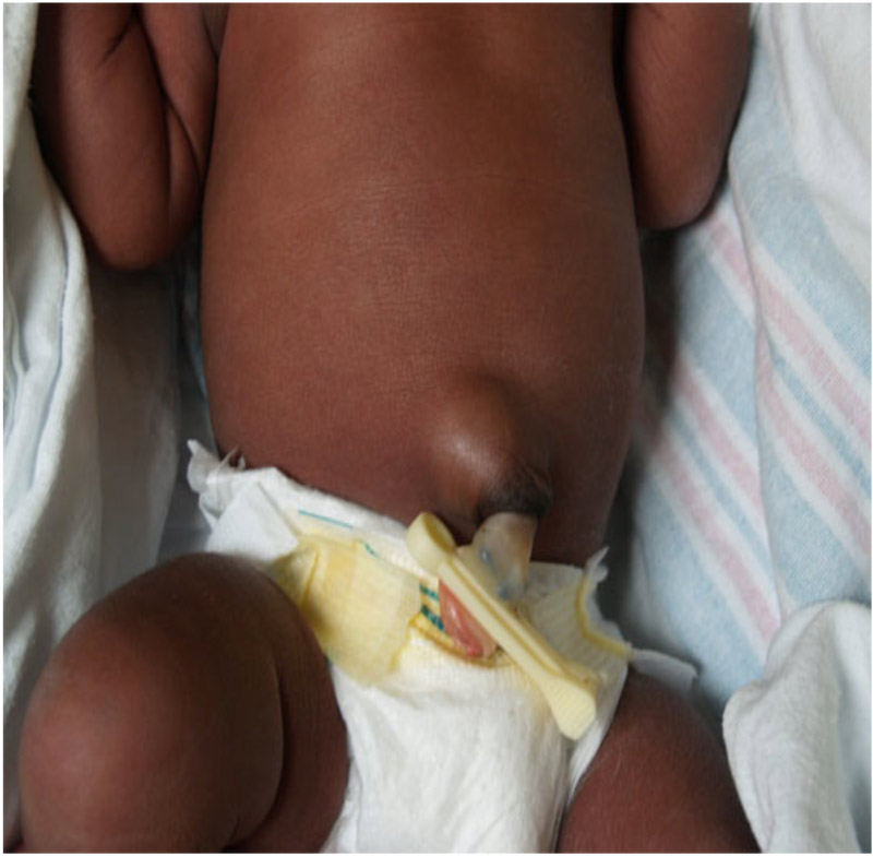
Umbilical hernias are common affecting 10% of Caucasian children25. The incidence is higher in preterm babies and those with Downs syndrome26.
Like the Diastasis Recti it presents as a painless, midline swelling in childs abdomen when intra-abdominal pressure is increased. However, in this case it is due to protrusion of the peritoneum-containing sac through the umbilical ring.
Incarceration is rare and overtime the ring constricts to obliterate the defect.
As spontaneous resolution is anticipated surgery is delayed until 4-6 years of age27 so most cases require reassurance only.
3) Communicating Hydrocoeles

Presents as a painless scrotal swelling due to collection of fluid inside the tunica vaginalis.
Occurs in neonates and infants occurs due to the presence of a persistent processus vaginalis where the testicle descends through the inguinal canal into the scrotum during foetal development.
It may be difficult to distinguish from a inguinal hernia but typically you can get above the swelling, no separate testicle can be felt and it trans-illuminates brightly. However, if there is uncertainty a Paediatric Surgeon should be consulted.
The persistent processus vaginalis will resolve spontaneously and hence, 89% will resolve in the first year of life28.
Most cases require reassurance only however, parents should be advised to return if symptoms of infection develop or there is a dramatic increase in size.
4) Undescended Testes

Most undescended testes are present at birth.
The condition affects 3-5% of term infants but can affect up to 30% of those who are premature29.
By 3 months the incidence falls to 0.8% and subsequent resolution is unlikely30. Hence, most cases will improve spontaneously and reassurance is all that is required.
Rational for treatment after 3 months is the prevention of testicular cancer, subfertility and testicular torsion. Hence, for older infants, outpatient surgical opinion is required.
NB: Bilateral non-palpable undescended testes in a phenotypical male should be considered a genetic female with congenital adrenal hyperplasia until proven otherwise
5) Umbilical Granulomas

Umbilical granulomas appear after cord separation and present as a soft, pink lesion with a moist appearance.
Treatment with silver nitrate is historically the gold standard however, chemical burns to the abdomen have been reported31.
There is evidence application of salt to the area led to 100% resolution of the condition without the inherent risk of the harm to the patient32
6) Prominent Xiphisterum

Presents are a firm, immobile lump in the midline of the chest.
Over time this normal anatomy will become less visible. Reassurance is all that is required.
Remember to work within your own competencies and follow local guidelines.
If the child appears unwell or you are unsure of the diagnosis you should always refer to your local friendly paediatrician or paediatric surgeon!
- Mason JK, Laurie GT. Mason and McCall Smiths Law and Medical Ethics. 7th edn. Oxford: Oxford University Press, 2006.
- Stewart MG. Evidence-based medicine in rhinology. Otolaryngology and Head and Neck Surgery 2008;16(1):14-17, February 2008.
- Newborn Photo Library. Stanford School of Medicine [Online]. Available from here [Accessed 28th February 2016].
- National Institute for Health and Care Excellence. Neonatal jaundice [CG98]. London: National Institute for Health and Care Excellence 2010.
- Batu ED, Yeni S, Teksam O. The factor affecting neonatal presentations to the pediatric emergency department. J Emerg Med 2015;48(5):542-7.
- Liu C, Feng J, Qu R, et al. Epidemiologic study of the predisposing factors in erythema toxicum neonatorum. Dermatology. 2005;210(4):269272.
- Berg FJ, Solomon LM. Erythemaneonatorum toxicum. Arch Dis Child 1987;62:327-328.
- Van Praag MC, Van Rooij RW, Folkers E, Spritzer R et al. Diagnosis and treatment of pustular disorders in the neonate. Pediatr Dermatol. 1997;14(2):131143.
- Schachner L, Press S. Vesicular, bullous and pustular disorders in infancy and childhood. Pediatr Clin North Am. 1983;30(4):609629.
- Laude TA. Approach to dermatologic disorders in black children. Semin Dermatol. 1995;14(1):1520.
- OConnor NR, McLaughlin MR, Ham P. Newborn skin: Part 1. Common Rashes. Am Fam Physician 2008;77(1):47-52.
- Selimoglu MA, Dilmen U, Karakelleoglu C, Bitlisli H, Tunnessen WW. Picture of the month. Harlequin color change. Arch Pediatr Adolesc Med. 1995;149(10):11711172.
- Williams ML. Differential diagnosis of seborrheic dermatitis. Pediatr Rev. 1986;7(7):204211.
- Tollesson A, Frithz A, Stenlund K. Malassezia furfur in infantile seborrheic dermatitis. Pediatr Dermatol. 1997;14(6):423425.
- Janniger CK. Infantile seborrheic dermatitis: an approach to cradle cap. Cutis. 1993;51(4):233235.
- Clinical Knowledge Summaries: seborrhoeic dermatitis (revised February 2013). Clinical Knowledge Summaries National Institute for Heath and Care Excellence [Online]. Available from: here [Accessed 23rd February 2016].
- National Institute for Health and Care Excellence. Neonatal jaundice [CG98]. London: National Institute for Health and Care Excellence 2010.
- National Institute of Child Health and Human Development randomized, controlled trials of phototherapy for neonatal hyperbilirubinemia. Pediatrics 1985; 75:(2 Pt 2)385-441.
- Sisson TR, Kendall N, Glauser SC et al. Phototherapy of jaundice in newborn infant. I. ABO blood group incompatibility. Journal of Pediatrics 1971;79:(6)904-10.
- Lewis HM, Campbell RH, Hambleton G. Use or abuse of phototherapy for physiological jaundice of newborn infants. Lancet 1982;2(8295)408-10.
- Meloni T, Costa S, Dore A et al. Phototherapy for neonatal hyperbilirubinemia in mature newborn infants with erythrocyte G-6- PD deficiency. Journal of Pediatrics 1974;85:(4)560-2.
- Martinez JC, Maisels MJ, Otheguy L et al. Hyperbilirubinemia in the breast-fed newborn: A controlled trial of four interventions. Pediatrics 1993;91:(2)470-3.
- Ju SH and Lin CH. The effect of moderate non-hemolytic jaundice and phototherapy on newborn behavior. Chung-Hua Min Kuo Hsiao Erh Ko i Hsueh Hui Tsa Chih 1991;32:(1)31-41.
- Morris BH, Oh W, Tyson JE et al. Aggressive vs. conservative phototherapy for infants with extremely low birth weight. New England Journal of Medicine 2008;359:(18)1885-96.
- Valdes OS, Maurer HM, Shumway CN et al. Controlled clinical trial of phenobarbital and-or light in reducing neonatal hyperbilirubinemia in a predominantly Negro population. Journal of Pediatrics 1971;79:(6)1015-7.
- Maurer HM, Shumway CN, Draper DA et al. Controlled trial comparing agar, intermittent phototherapy, and continuous phototherapy for reducing neonatal hyperbilirubinemia. Journal of Pediatrics 1973;82:(1)73-6.
- Wu PY, Lim RC, Hodgman JE et al. Effect of phototherapy in preterm infants on growth in the neonatal period. Journal of Pediatrics 1974;85:(4)563-6.
- Curtis-Cohen M, Stahl GE, Costarino AT et al. Randomized trial of prophylactic phototherapy in the infant with very low birth weight. Journal of Pediatrics 1985;107:(1)121-4.
- Disorders of the umbilicus in infants and children: A consensus statement of the Canadian Association of Paediatric Surgeons. Paediatrics and Child Health 2001;6(6):312-313.
- ODonnell KA, Glick PL, Caty MG. Pediatric umbilical problems. Pediatr Clin North Am 1998;45:7919.
- Skinner MA, Grosfeld JL. Inguinal and umbilical hernia repair in infants and children. Surg Clin North Am 1993;73:43949.
- Naji H, Ingolfsson I, Isacson D, Svensson JF. Decision making in the management of hydroceles in infants and children. Eur J Pediatr 2012;171(5):807-10.
- Docimo SG, Silver RI, Cromie W. The undescended testicle: diagnosis and management. Am Fam Physician 2000;62(9):2037-44, 2074-8.
- Berkowitz GS, Lapinski RH, Dolgin SE, Gazella JG, Bodian CA, Holzman IR. Prevalence and natural history of cryptorchidism. Pediatrics 1993;92:449.
- Chamberlain JM, Gorman RL, Young GM. Silver nitrate burns following treatment for umbilical granuloma. Pediatr emerg care 1992;8(1):29-30.
- Kesaree N, Babu PS, Banapurmath CR, Krishnamurthy SN. Umbilical granuloma. Indian Pediatrics 1983;20(9):690-2.